.webp)
In collaboration with Surgical Data Science Collective (SDSC), researchers at Stanford University Department of Neurosurgery presented their cutting-edge research at the 2024 American Rhinologic Society (ARS) Meeting. It was an incredible opportunity to showcase their innovative work facilitated by our Surgical Video Platform (SVP), where they used SDSC’s Artificial Intelligence (AI) models to study surgical technique in a variety of endoscopic-based neurosurgical procedures.
In the world of endoscopic surgery, precision and technique are paramount, especially for challenging cases of brain tumor resection. SDSC is revolutionizing the surgical field with AI tools that analyze surgical video footage to improve understanding, training, and clinical outcomes through the Surgical Video Platform (SVP). Leveraging deep machine learning (ML) models, the SVP’s capabilities extend beyond traditional metrics, delivering a comprehensive look at instrument usage, technique patterns, and procedural steps that are essential in complex surgeries. This pioneering approach opens doors to new levels of surgical precision, enhanced training, technique standardization, and ultimately, patient outcomes.
There are a plethora of ways in which the SVP can be harnessed for research, including the use of AI to clarify and delineate the nuances of surgical technique, and progress towards an objective measure of surgeon performance. The SVP’s cutting-edge metric analysis tools can be adapted to a handful of endoscopic or microscopic procedures, opening new avenues for surgical advancement and clinical research across any operative discipline.
Here are some of the neurosurgical cases Stanford University showcased at ARS 2024:
Pituitary adenoma with transsphenoidal resection (TSSR)
David R. Grimm MD1, Dhiraj Pangal MD1, Danyal Z. Khan MD1, Margaux Masson-Forsythe, Ayesha Syeda, Jack Cook, Peter H. Hwang MD1, Jayakar Nayak MD PhD1, Zara M. Patel MD1, Noel Ayoub MD MBA1, Daniel Donoho MD, Juan Fernandez-Miranda MD1, Michael T. Chang MD1
1Department of Otolaryngology-Head and Neck Surgery, Stanford University School of Medicine
Pituitary adenomas (PA) are a type of benign brain tumor that arise in the pituitary gland. This gland is responsible for making, storing, and releasing hormones into the body. They are typically removed by endoscopic endonasal transsphenoidal surgery, also called an adenomectomy, where the adenoma is removed through the nose and sinuses.
SDSC developed a YoloV8-based neural AI network for instrument recognition and spatiotemporal tracking, allowing researchers at Stanford University to examine footage for the identification of conserved tool motion patterns during endonasal skull base surgery. In a sample of five patients, Stanford generated instrument motion plots using tool detection prediction CSV files exported from the SVP. Heatmaps were also generated for beginning-to-end statistical video analysis of the procedures.

Key findings included patterns in common instruments used by surgical stage, quantified with the Dice-Sørensen Similarity Coefficient (DSC), a metric for assessing similarity in tool motion across different cases. Stanford also used SVP-generated results to highlight that several instrument motion patterns are more highly conserved in initial surgical phases compared to later ones. Although embryonic, this pilot study demonstrates a potential framework for evaluating surgical performance utilizing AI-driven automated video analysis, and drives us closer to drawing clinical correlations between techniques in the future.
Uncovering Nuanced Techniques in Subarachnoid Invasion Cases
Jonathan B. Lamano, MD/PhD1, Ana S. Alvarez, MD1, Dhiraj J. Pangal, MD1, Juan C. Fernandez-Miranda, MD1
1Department of Neurosurgery, Stanford University, Stanford, California
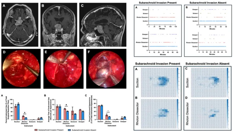
Certain cases of PA involve tackling an additional complication of subarachnoid invasion, where the tumor has encroached on vital neurovascular structures. This study applied SDSC’s ML modules to quantitatively assess the modified technique that surgeons use to avoid damage to these vulnerable areas.
By analyzing instrument timelines, usage statistics, and heatmaps, the SVP identified increases in key tool frequencies and use duration in cases where subarachnoid invasion was present. Prolonged instrument use and spatial tracking indicated critical precision-intensive parts of the surgery, as well as heatmaps demonstrating the careful maneuvering of tools around delicate structures.

Overall, this study demonstrates how the SVP is facilitating the initial steps to quantitatively and qualitatively characterize the nuances in surgical approach. AI-derived insights are invaluable in creating refined guidelines for the surgical intervention of complex pathology, and are transforming the future of surgical safety and efficacy.
Defining Resection Techniques for Cavernous Sinus Invasion
Jonathan B. Lamano, MD/PhD1, A. Sofia Alvarez, MD1, Dhiraj J. Pangal, MD1, Juan C. Fernandez-Miranda, MD1
1Department of Neurosurgery, Stanford University, Stanford, California
Pituitary adenomas that invade the cavernous sinus also offer a unique surgical challenge. Incomplete resection presents a significant risk of recurrence or lack of remission, and so researchers at Stanford University are attempting to characterize the most safe and effective surgical technique adopting the same SVP resources as the previous study.

Here, SVP ML models were applied to a cohort of nine patients, and harnessed to identify divergence in instrument use between the sellar and transcavernous components of the surgery. The suction and Rhoton dissector were most heavily used during the transcavernous phase, underscoring the role of controlled tissue manipulation and bleeding management.
Evaluating surgical technique from both start-to-finish, and breaking it down into procedural steps, allows surgeons to extract as many learning opportunities as possible from every operation. Employing AI for the creation of refined guidelines can help surgeons avoid damage to compromised structures, as well as provide a unique educational opportunity for aspiring surgeons to dissect their practices, and improve surgical success for their patients.
Redefining Surgical Performance with AI
SDSC’s surgical research approach stresses the value of building a global repository of surgical video data. By accumulating insights from varied cases and techniques, SDSC envisions a database that supports surgeons worldwide in understanding the best practices, assessing performance, and refining techniques over time. AI-generated insights into instrument movement patterns, procedural phases, and technique-specific demands has the potential to enhance both surgical education and intraoperative decision-making.
Moreover, SDSC’s work sets a foundation for developing standardized, objective performance metrics that could be incorporated into training programs, offering an unprecedented, data-driven pathway for assessing and improving surgical skills.
This structured approach enables both experienced surgeons and trainees to understand not only “how” procedures are performed, but “why” certain techniques are superior in specific anatomical or pathological scenarios. As the SVP continues to evolve, it seeks to support a new paradigm of data-driven surgical assessment, enhance training methods, and ultimately, contribute to better patient outcomes across the field of endoscopic surgery.
SDSC invites you to join our platform and collaborate with our brilliant team to drive your research initiatives forward!


